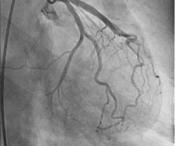
The transfer of patients with a suspected myocardial infarction (MI) from hospitals without catheterization laboratories (cath labs) to those equipped with such facilities is a critical process that demands precise planning and execution. This comprehensive guide outlines the necessary steps, medications, and protocols to ensure patient stability and safety during this crucial period.
Initial Assessment and Stabilization
Patient History and Symptoms
- Obtain a Detailed History: Gather information about the onset, duration, and characteristics of chest pain. Ask about associated symptoms such as shortness of breath, sweating, nausea, or lightheadedness.
- Identify Risk Factors: Note risk factors for coronary artery disease, including hypertension, diabetes, smoking, family history, hyperlipidemia, and previous cardiovascular events.
Physical Examination
- Vital Signs Monitoring: Record blood pressure, heart rate, respiratory rate, and oxygen saturation.
- Cardiac Examination: Listen for heart murmurs, extra heart sounds (S3, S4), and jugular venous distention.
- Respiratory Assessment: Check for signs of pulmonary congestion or edema.
- Peripheral Assessment: Observe for signs of poor perfusion, such as cold extremities, pallor, or cyanosis.
Electrocardiogram (ECG)
- Perform a 12-Lead ECG: This is the primary diagnostic tool to identify ischemic changes, ST-segment elevation, T-wave inversions, or new left bundle branch block (LBBB), which indicate an acute MI.
- Continuous Monitoring: Place the patient on continuous ECG monitoring to detect any arrhythmias or changes in the ST segment.
Laboratory Tests
- Cardiac Biomarkers: Obtain blood samples to measure troponin levels, which are specific markers for myocardial injury.
- Complete Blood Count (CBC): Check for anemia or signs of infection.
- Basic Metabolic Panel (BMP): Assess kidney function, electrolytes, and glucose levels.
- Coagulation Profile: Evaluate clotting status, especially if anticoagulation therapy is being considered.
Immediate Stabilization
Oxygen Therapy
- Administer Supplemental Oxygen: Provide oxygen if the patient is hypoxic (SpO2 < 94%) to improve tissue oxygenation.
Medication Administration
- Aspirin: Administer chewable aspirin (162-325 mg) immediately to inhibit platelet aggregation.
- Nitroglycerin: Give sublingual or IV nitroglycerin to relieve chest pain and reduce myocardial oxygen demand. Monitor blood pressure closely to avoid hypotension.
- Morphine: Use morphine for pain relief and to reduce anxiety, which can decrease the heart’s oxygen demand. Be cautious with dosing and monitor for adverse effects like hypotension and respiratory depression.
- Beta-Blockers: Administer if not contraindicated (e.g., metoprolol) to reduce heart rate and myocardial oxygen consumption.
- Heparin: Start anticoagulation therapy with unfractionated or low molecular weight heparin to prevent further clot formation.
- Clopidogrel or Other P2Y12 Inhibitors: Consider as the patient is to undergo percutaneous coronary intervention (PCI).
Advanced Cardiac Life Support (ACLS) Protocols
- Defibrillation/Cardioversion: Be prepared to perform defibrillation or synchronized cardioversion in case of life-threatening arrhythmias.
- CPR: Initiate cardiopulmonary resuscitation if the patient goes into cardiac arrest.
Preparation for Transfer
Stabilization of Vital Signs
- Ensure Stable Hemodynamics: Maintain systolic blood pressure above 90 mmHg and adequate perfusion.
- Control Heart Rate: Aim for a heart rate between 60-100 bpm, unless contraindicated by the patient’s condition.
Prophylactic Measures
- Allergy Prevention: Administer A1 histamine blocker (e.g., diphenhydramine) and H2 histamine blocker (e.g., ranitidine) for patients with known contrast dye allergies.
- Corticosteroids: Give methylprednisolone (Solu-Medrol) to reduce the risk of severe allergic reactions to contrast dye.
Communication and Documentation
- Prepare a Detailed Transfer Report: Include patient history, treatments administered, vital signs, ECG findings, and any complications.
- Coordinate with Receiving Facility: Ensure the cath lab team is ready and aware of the patient’s condition and estimated time of arrival.
Transport Arrangements
- Select Appropriate Transport: Use an ambulance equipped with advanced life support (ALS) capabilities.
- Escort by Trained Personnel: Ensure the presence of a critical care paramedic and/or nurse trained in cardiac care during transport.
Additional Medications for Comprehensive Management
- Morphine: For pain relief and to reduce anxiety.
- Beta-Blockers (e.g., Metoprolol, Atenolol): To manage high blood pressure and reduce heart rate.
- Heparin (Unfractionated or Low Molecular Weight): To prevent further clot formation.
- Oxygen Therapy: To improve oxygenation in patients with low blood oxygen levels.
- Clopidogrel or Other P2Y12 Inhibitors (e.g., Prasugrel, Ticagrelor): To prevent further platelet aggregation and clot formation.
- Statins (e.g., Atorvastatin, Rosuvastatin): To stabilize atherosclerotic plaques and reduce lipid levels.
- Calcium Channel Blockers (e.g., Diltiazem, Verapamil): To manage hypertension and angina.
- Furosemide (Lasix): To manage fluid overload.
- Magnesium Sulfate: To manage arrhythmias or prevent complications from low magnesium levels.
- ACE Inhibitors (e.g., Lisinopril, Enalapril): To manage hypertension and reduce heart workload.
Conclusion
The safe transfer of patients with suspected myocardial infarction to facilities with cath labs involves meticulous assessment, stabilization, and coordination. By following a systematic approach to diagnosing and managing MI, administering essential medications, and preparing for potential complications, healthcare providers can ensure optimal patient outcomes. Effective communication and coordination between the sending and receiving facilities are crucial to facilitate a smooth transfer process, ultimately improving the chances of successful patient care and recovery.
References
- American Heart Association. (2020). “Managing Patients with Acute Myocardial Infarction: Guidelines for Emergency Medical Services”. Retrieved from American Heart Association.
- National Institute for Health and Care Excellence (NICE). (2021). “Acute Coronary Syndromes: Initial Assessment and Treatment”. Retrieved from NICE.
- European Society of Cardiology (ESC). (2020). “ESC Guidelines for the Management of Acute Myocardial Infarction in Patients Presenting with ST-segment Elevation”. Retrieved from ESC.
- Mayo Clinic. (2021). “Allergic Reactions to Contrast Dye”. Retrieved from Mayo Clinic.
- U.S. National Library of Medicine. (2020). “Histamine Blockers: Uses, Side Effects, and Precautions”. Retrieved from MedlinePlus.
- Journal of the American Medical Association (JAMA). (2019). “Corticosteroids in the Prevention of Allergic Reactions to Contrast Dye”. JAMA, 322(15), 1463-1471.
- American College of Cardiology (ACC). (2021). “Prehospital Management of Acute Coronary Syndrome”. Retrieved from ACC.


